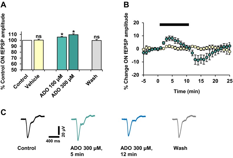
Fig. 4.
The effect of adenosine on the NMDAR-mediated RGC ON-fEPSP. Adenosine (ADO) was applied (10 min) at concentrations of 100 and 300 μM. n = 8 for both concentrations tested. a Values are mean ± SEM percentage of pre-treatment control fEPSP peak amplitude, taken ∼5–7 min after the start of adenosine application. The ON-fEPSP was reversibly and significantly potentiated by adenosine, in a concentration-related manner. Vehicle application elicited no effect on the ON-fEPSP. ns not significant; *P < 0.05, compared to control. b Time course plot shows the effect of adenosine (300 μM; green) and vehicle (yellow) on the ON-fEPSP. Plotted values are mean ± SEM percentage change in fEPSP peak amplitude relative to pre-treatment control. c Representative traces (120 s averages) illustrate the effect of adenosine (300 μM) on the ON-fEPSPs

