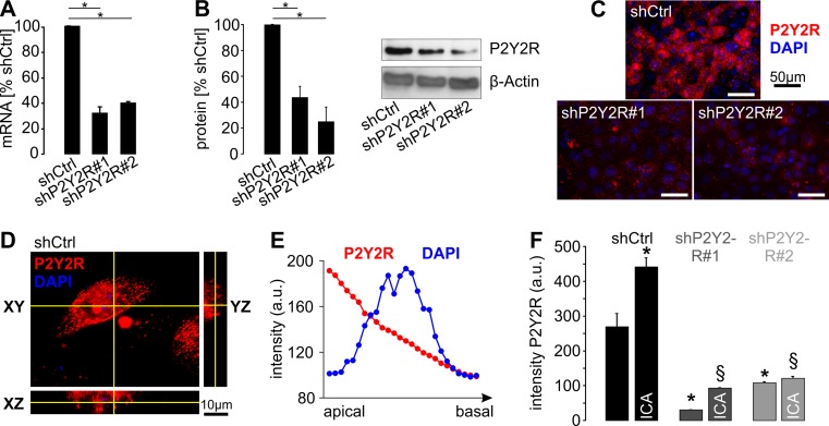
Fig. 3.
P2Y2R is localized in the apical membrane of plMDCK cells, and its expression is regulated by HIF-1α. a Quantification of P2Y2R mRNA in plMDCK cell clones stably transfected with two distinct shRNAs directed against P2Y2R (shP2Y2R#1 and shP2Y2R#2) compared with MDCK cells stably transfected with scrambled shRNA (shCtrl) serving as control (n = 4). b Quantification of P2Y2R protein expression in P2Y2R knockdown and control cells described in a (n = 5). Right panel shows representative Western blot. c Representative photos of plMDCK cells competent (shCtrl) or deficient for P2Y2R (shP2Y2R#1 and #2) that were grown on permeable supports and stained for P2Y2R (red) and nuclei (DAPI; blue). d Representative z-stack analysis of plMDCK cells stained for P2Y2R (red) and DAPI (blue) in control-transfected, polarized plMDCK cells showing P2Y2R signals predominantly at the top of the cells and much less pronounced at the bottom. e Representative quantification of P2Y2R signal (red) and DAPI (blue) along the z-axis from the apical to the basal side of the cell. f Quantification of the apical P2Y2R signal intensity in P2Y2R competent (shCtrl) and deficient (shP2Y2R#1 and #2) plMDCK cells in the absence and presence of 10 μM ICA. Asterisk indicates comparison to non-treated control-transfected cells. Section sign indicates comparison to ICA-treated control-transfected cells

