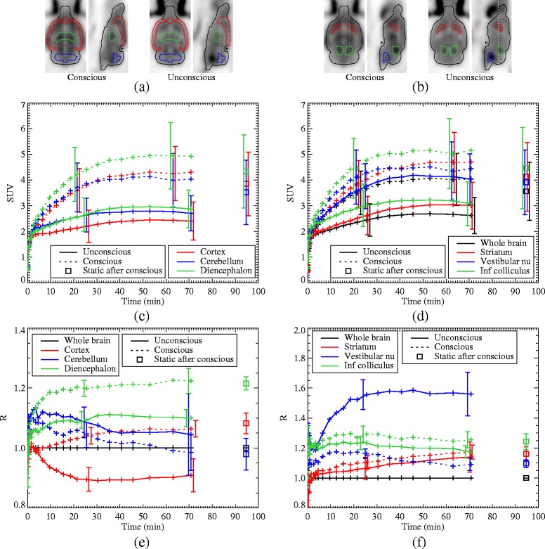
Fig. 6.

a, b The ROIs under consideration are indicated on the last frame from the average of the eight reconstructions. c, d TACs for various ROIs for the average of the eight data sets, for the conscious (dotted lines) and unconscious (solid lines) scans, as well as the static scan with anaesthesia following the conscious scan (square points). The overlapping error bars are staggered for clarity. e, f The ratio of various ROIs to the whole brain average, averaged over the eight data sets. In all plots, the error bars are the standard deviations across the data sets and are only shown in two representative locations for clarity
