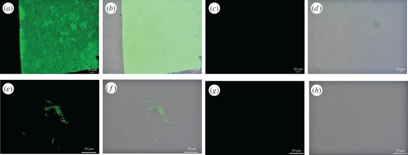
Figure 5.
Immunohistochemical staining results for (a–d) ostrich claw sheath and (e–h) Citipati claw sheath exposed to antiserum raised against avian feathers [33]; (a, c, e and g) are imaged using a FITC filter; (b, d, f and h) are overlay images, superimposing fluorescent signal on a transmitted light image of sectioned tissue to reveal the localization of antibody–antigen complexes to tissue. Positive binding is observed in both extant and fossil claw sheath material. Controls using secondary antibody only are negative for both (c,d) ostrich and (g,h) fossil material. (Online version in colour.)

