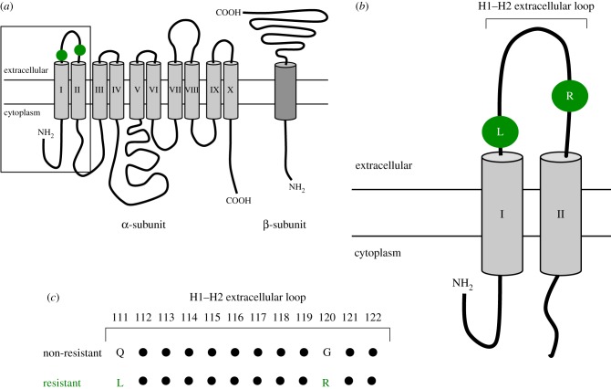
Figure 1.
(a) Schematic diagram of an unfolded Na+/K+-ATPase molecule, showing the location of the Q111 L and G120R substitutions on the H1–H2 extracellular loop. The box shows the area enlarged in b. (b) Enlargement of the H1–H2 extracellular loop and the first two transmembrane domains. (c) The amino acid sequence of the first extracellular loop, indicating the substitutions at positions 111 and 120 that confer resistance to bufadienolides in snakes. Redrawn from Köksoy [21]. For a more detailed structural reconstruction of the interactions of the Na+/K+-ATPase and a BD, see Laursen et al. [22]. (Online version in colour.)

