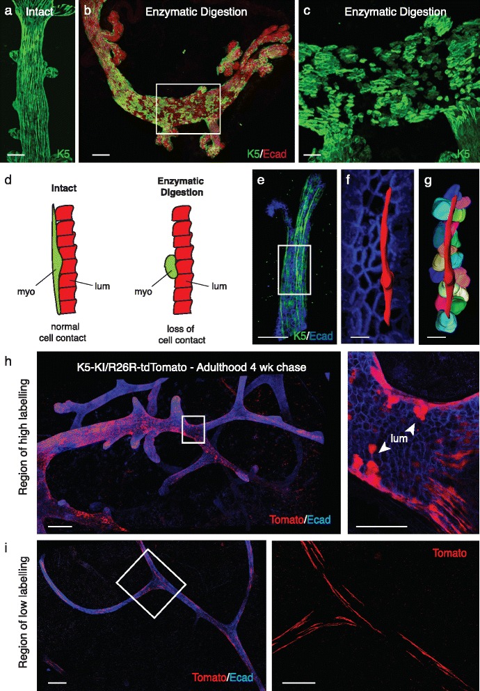
Fig. 1.

Influence of whole-mount preparation and efficiency of labelling on lineage tracing outcomes. a Whole-mount three-dimensional (3D) confocal image of an intact ductal portion immunostained for Keratin 5 obtained using a protocol that does not include any enzymatic digestion (described in Rios et al. [4]). Scale bar = 50 μm. b Whole-mount 3D confocal image of a ductal portion (8-week-old FVB/N mouse) immunostained for Keratin 5 (green) and E-cadherin (red), using the protocol described by Wuidart et al. for enzymatic digestion of mammary glands [1]. The mammary portions were subsequently fixed in 4% paraformaldehyde (PFA) for 2 h at 4 °C, and processed as described in Rios et al. [4] for immunostaining and confocal imaging. c Image of an enlargement from (b) showing the myoepithelial layer immunostained for Keratin 5. Note the paucity of myoepithelial cells and their altered shape, which is no longer elongated. Scale bars = 100 μm (b) and 30 μm (c). d Schematic panels showing normal contacts between one myoepithelial cell (myo, in green) and multiple luminal cells (lum, in red) in intact breast tissue (left), and loss of cell-cell contacts between the myoepithelial (in green) and luminal cells (in red) after enzymatic digestion (right). e Whole-mount 3D confocal image (from Fig. 1d, Rios et al. [4]) of a duct in a K5rtTA-IRES-GFP mammary gland immunostained for E-cadherin (in blue). f enlargement from (e), showing a mask (in red) applied to one myoepithelial cell (GFP channel using Imaris software) and the luminal layer labelled with E-cadherin (in blue). g Image showing the mask of the myoepithelial cell and the masks of all luminal cells in direct contact with the myoepithelial cell. Note: the mask was applied to a representative 70 μm myoepithelial cell. Scale bars = 50 μm (whole-mount e) and 10 μm (enlargements f, g). h, i Whole-mount 3D confocal images of ducts in a K5-KI/R26R-tdTomato mammary gland, showing either a high (h) or low (i) degree of labelling in the same gland, at 4 weeks post-induction with tamoxifen in adulthood and then immunostained for E-cadherin (blue). Enlargements show optical sections depicting luminal cell-containing clones (upper panel) or elongated myoepithelial cells (lower panel). Scale bars = 200 μm (whole-mounts) and 50 μm (enlargements)
