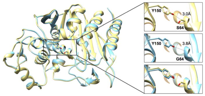
Figure 3.
Structure alignment of P99 wild type and S64G mutant β-lactamases. The wild-type P99 structure (PDB ID: 1XX2) is shown in yellow and the P99 S64G mutant structure determined in this study is shown in cyan. The inset contains a detailed view of residues 64 and 150. The absence of the side chain at position 64 in the S64G mutant (middle panel) increases the distance between Oη of the Y150 and position 64 to 3.8 Å compared to 3.0 Å in the wild type (top panel).

