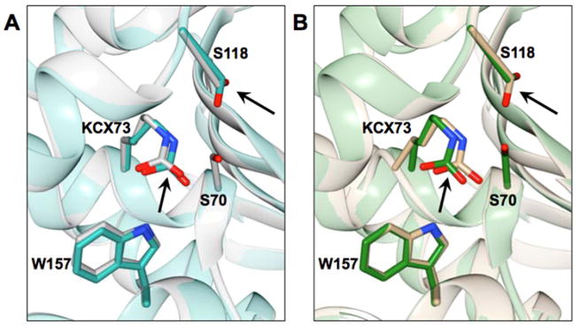
Figure 5.
The position of Lys73 is shown in the crystal structure of the S70G mutant of OXA-48 (dark cyan) aligned with wild-type OXA-48 (PDB ID: 3HBR) in grey (left panel). The right panel shows the S70G mutant of OXA-163 in tan aligned with OXA-163 (PDB ID: 4S2L) in green. Selected residues are represented in stick and labeled. Black arrows point at the Lys73 and Ser118 residues outlining the movement of these residues in the S70G mutants in comparison to the wild-type enzymes.

