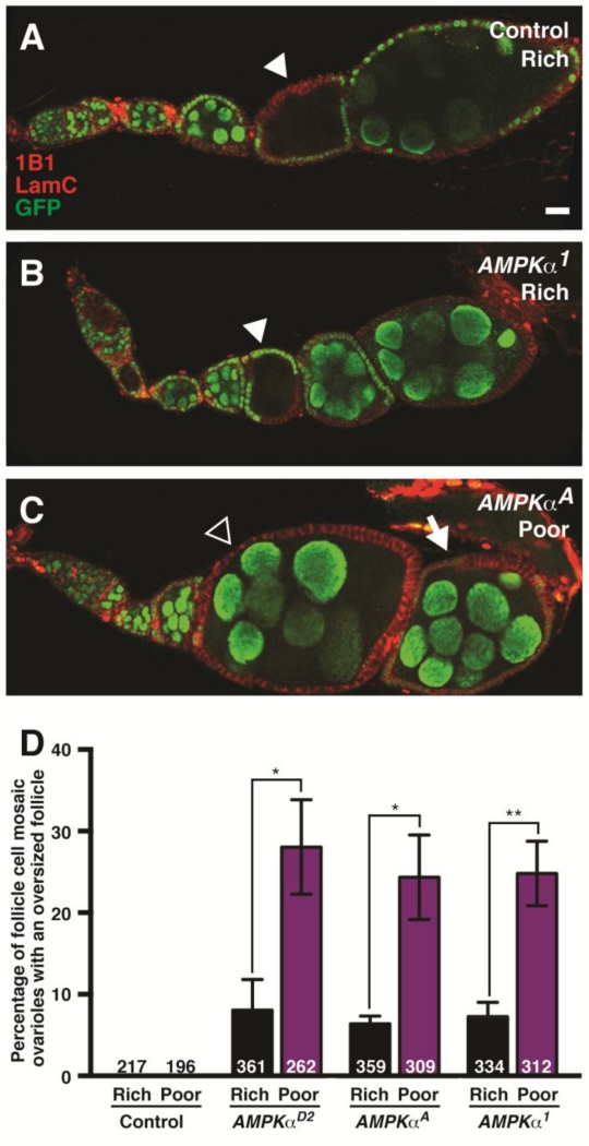
Figure 3. AMPK function is required in follicle cells, but not in the germline, for follicle growth.
(A and B) Control (A) and AMPKα mutant mosaic (B) ovarioles with GFP-negative germline cysts (arrowheads) that grow normally relative to flanking GFP-positive cysts. (C) AMPKα mutant mosaic ovariole showing overgrowth of a follicle containing a wild-type germline cyst surrounded by AMPKα mutant follicle cells (open arrowhead) relative to the posterior, older follicle, which contains fewer GFP-negative follicle cells (arrow). GFP (green) labels wild-type cell nuclei; 1B1 (red) labels fusomes and cell membranes; Lamin C (LamC; red) labels cap cell nuclear envelopes. Scale bar, 10 μm. (D) Quantification of follicle overgrowth in follicle cell mosaic ovarioles at 7 days after clone induction. This phenotype is markedly enhanced on poor relative to rich diets. Sample sizes are shown inside bars and represent results from three independent experiments. Error bars represent S.E.M. *p<0.05; **p<0.01 by Student’s t test.

