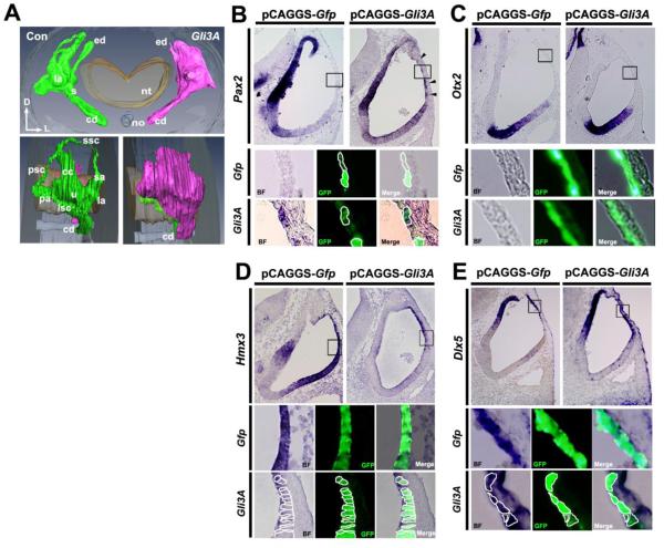
Figure 5. Altering the GLI3R/GLI3FL ratio by overexpressing Gli3A in the dorsolateral otocyst perturbs DV polarity of the otocyst.
(A) Reconstruction of serial section of chick inner ears collected 5-6 days after Gli3A overexpression (as shown in Fig. 3A). Frontal (top single panel) and lateral (bottom paired panels) are shown. n=5 embryos sectioned; n=1 embryo reconstructed. (B-E) ISH of sections of chick otocysts collected 12-16 hours after overexpression of Gfp alone or Gli3A and Gfp showing Pax2, Otx2, Hmx3, and Dlx5 expression. For Otx2: n=3 for both control and experimental embryos; for Pax2, Hmx3, and Dlx5: n=4 for both control and experimental embryos. Boxes show regions enlarged below, with borders of selected cells outlined. Arrowheads in B indicate dorsal otocyst cells ectopically expressing Pax2.

