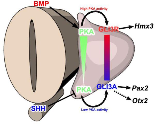
Figure 6. Model: PKA activity is a common intersection point coordinating dorsalizing and ventralizing signals in the early otocyst.
SHH signaling emanating from the floor plate of the neural tube and notochord maintains low PKA activity in the ventral otocyst, whereas BMP signaling in the dorsal otocyst activates PKA; thus, a gradient of PKA activity forms across the DV axis of the otocyst. High PKA activity in the dorsal otocyst results in high GLI3FL partial proteolytic processing, the accumulation of GLI3R, and the expression of Hmx3. In contrast, low PKA activity in the ventral otocyst results in low GLI3FL proteolytic processing, the maintenance of GLI3FL (potentially GLI3 activator), and the expression of Pax2 and Otx2.

