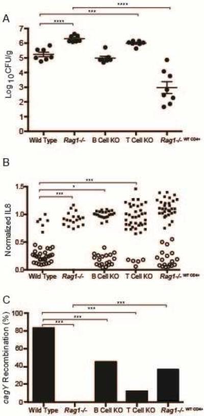
Figure 1. CD4+ T cells are required to control H pylori colonization density and select strains with loss of T4SS function and recombination in cagY.
(A) H pylori colonization density was significantly greater in Rag1−/− and T cell KO mice than in wild type mice. Adoptive transfer of WT CD4+ T cells into Rag1−/− mice markedly reduced H pylori colonization compared to Rag1−/−. Each data point represents CFU/g for an individual mouse 8 weeks PI (N=7–8 mice/group). Horizontal lines indicate mean ± standard error of the mean (SEM). (B) Single colonies recovered from WT and B cell KO mice, and mice adoptively transferred with WT CD4+ T cells, showed marked loss in the capacity to induce IL8 that was accompanied by recombination in cagY (open circles). In contrast, all colonies from Rag1−/− mice and most from T cell KO mice induced IL8 and had the same cagY RFLP (closed circles) as WT H pylori PMSS1. Each data point represents the result from a single colony (N=3-6 colonies/mouse). (C) Percent of colonies that underwent cagY recombination (open circles divided by total colonies for each group in panel B). *P≤0.05, **P≤0.01, ***P≤0.001.

