Figure 1.
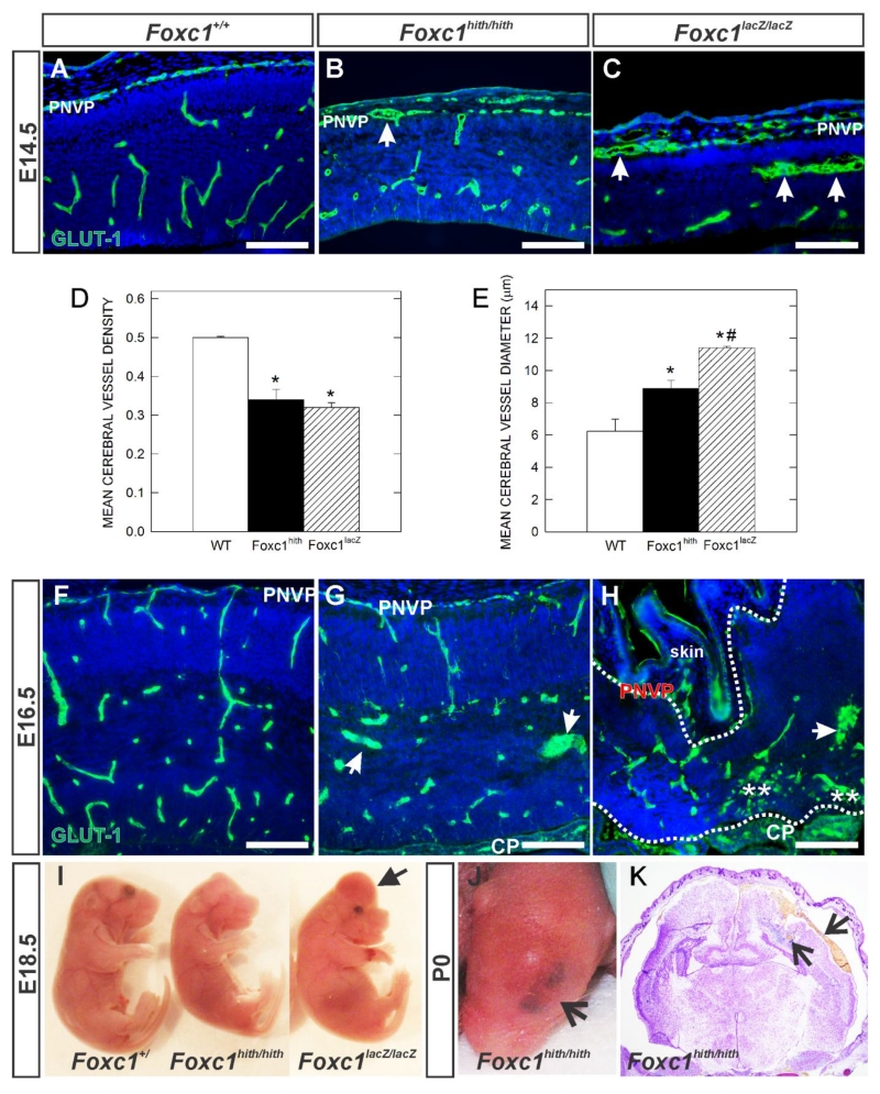
(A-C, F-H) Immunohistochemistry of E14.5 and E16.5 neocortex of wildtype (Foxc1+/+) and Foxc1 mutants (Foxc1hith/hith and Foxc1lacZ/lacZ) labeled with GLUT1, a CNS blood vessel marker and DAPI. Arrows indicate enlarged, dysplastic vessels. PNVP: Peri-neural vascular plexus. (D, E) Graphs depict quantification of average vessel density (n=3) and vessel diameter (n=4) of E14.5 cerebral vasculature in wildtype (WT), Foxc1 hypomorph (Foxc1hith) and knockout (Foxc1lacZ) animals. * indicate significant difference from wildtype (p<0.05) and *# indicates significant difference from wildtype and Foxc1hith (p<0.05). (I) E18.5 embryos harvested from wildtype and Foxc1 mutants showing forebrain hemorrhage in Foxc1lacZ/lacZ mutant. (J) Postnatal day (P) 0 Foxc1hith/hith mutant showing a characteristic bruise in the head. (K) Foxc1hith/hith coronal section of the brain showing hemorrhage in cerebral cortex (arrow). Scale bars = 100 μm.

