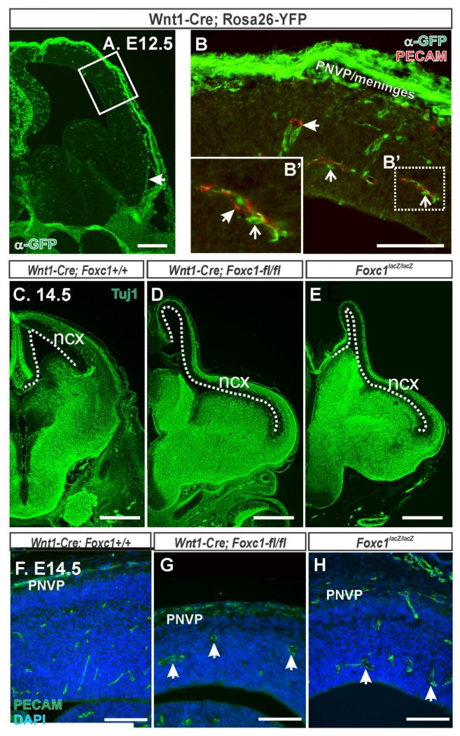
Figure 3.
(A) Low magnification image at the level of the telencephalon of E12.5 Wnt1-Cre; Rosa26-YFP, YFP signal (green) indicates Wnt1-Cre recombined cells. Box highlights area shown at higher magnification in B. (B) Higher magnification image of the neocortex and overlaying mesenchyme in an E12.5 Wnt1-Cre; Rosa26-YFP immunolabeled with PECAM (red). YFP+ cells were apparent in the PNVP and meninges. Closed arrows depict PECAM+/YFP− blood vessel and open arrows depict YFP+ perivascular pericytes in B and B’. E14.5 embryonic brains from (C) Wnt1-Cre;Foxc1+/+, (D) Wnt1-Cre;Foxc1-fl/fl and (E) Foxc1lacZ/lacZ immunostained with Tuj1 (green) which labels young neurons. Dashed line outlines length of neocortex (ncx). E14.5 neocortex from (F) Wnt1-Cre;Foxc1+/+, (G) Wnt1-Cre;Foxc1-fl/fl, and (H) Foxc1lacZ/lacZ immunolabled with PECAM. Arrows in G, H indicate dilated cerebral vessels. Scale bars = 200 μm (A), 500 μm (C-E), 100 μm (B,F-H).

