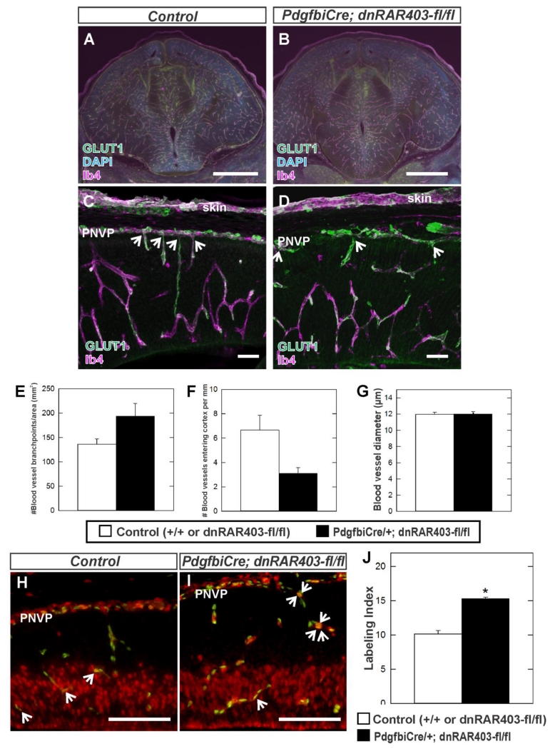
Figure 5.
(A-D) Immunohistochemistry of E14.5 coronal brain sections at the level of the forebrain (A, B) and in the neocortex (C, D) labeled with GLUT1 (green) and IB4 (cyan) from control and Pdgfbi-Cre;dnRAR403-fl/fl mutant samples. (E-G) Quantification of vessel branch points, number of blood vessels growing into the cortex from the PNVP and vessel diameter in control (n=3) and Pdgfbi-Cre;dnRAR403-fl/fl (n=3). Arrows in C and D indicate PNVP-derived blood vessels. Asterisks indicate statistically significant difference (p<0.05) from control. Immunohistochemistry of E14.5 cerebral cortex from (H) control and (I) Pdgfbi-Cre; dnRAR403-fl/fl mutant labeled with endothelial cell nuclear marker ERG1 (green) and BrdU (red) which labels cells in S phase. Arrows indicate ERG1+/BrdU+cells. (J) Quantification of endothelial cell proliferation (Labeling Index) in control (n=3) and Pdgfbi-Cre;dnRAR403-fl/fl mutants (n=3). Asterisk indicates statistically significant difference (p<0.05) from control. Scale bars = 1000 μm (A-B), 50 μm (C, D), 100 μm (H, I).

