
Figure 1. Comparison between synthetic (GUVs) and biological (GPMVs) membrane model systems.
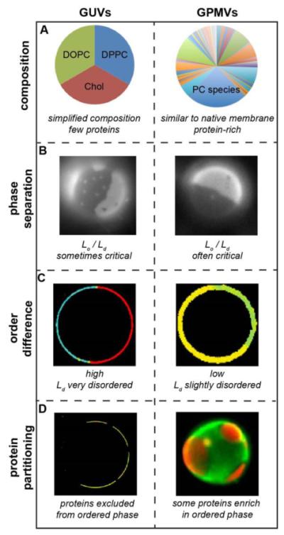
(A) Synthetic and biological model membranes differ dramatically with respect to their lipid and protein composition. Synthetic model membranes like GUVs are typically composed of a small number of different lipid components and few, if any, proteins. Shown is a canonical “raft mixture” containing equimolar saturated (DPPC) and unsaturated (DOPC) phospholipid, and cholesterol (Chol). In contrast, biological membrane models like GPMVs contain hundreds of different lipids and likely thousands of proteins. Shown is a representative distribution of phosphatidylcholine species in a PM sample, which contains a few major species (e.g. POPC is ~25 mol% of all PC) and over 100 minor ones54. (B) Despite these differences, GUVs and GPMVs are morphologically similar and both separate into coexisting ordered and disordered phases, visualized by staining vesicles with lipidic dyes that preferentially partitioning into one of the phases93. Notably, GPMVs often show near-critical behavior, also achievable in GUVs at specific compositions. (C) Despite these superficial similarities, these are notable difference in domain properties between GPMVs and GUVs. For example, coexisting domains in GPMVs are much more similar than in GUVs with respect to lipid packing. Images shown are pseudocolored maps of Laurdan generalized polarization, a widely used proxy for lipid packing (for experimental details, see references18, 160). (D) The relatively high disparity possibly results in most components being excluded from GUV ordered domains, whereas a number of lipids and proteins partition preferentially to this phase in GPMVs. Images show the partition of a peptide based on the transmembrane protein LAT. Yellow pseudocolor represents co-partitioning with a disordered phase marker in GUVs, while distinct red staining of one phase versus green in the other shows that the LAT-RFP protein partitions away from the disordered phase marker in GPMVs (for details, see references46, 57).