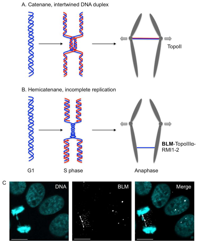
Fig. 4.
Model for the origin of ultra-fine bridges (UFBs). (A) Fully replicated but still intertwined DNA duplexes give rise to UFBs in anaphase and are resolved by topoisomerase II. These catenanes structures are commonly observed at centromeres. (B) Late replication intermediates lead to UFBs in anaphase. These hemicatenane structures arise predominantly from fragile sites under replicative stress and are resolved by BLM-TopoIII -RMI1-2. Parental DNA molecule is depicted in blue and in red is the nascent strand synthesized in S phase (C) Visualization of UFB in anaphase. HeLa cells were fixed and stained for BLM (white) and DAPI (cyan). In interphase, BLM localizes to distinct nuclear loci (PML nuclear bodies) seen as discreet foci. In anaphase, BLM localizes to UFB that appears as a thin thread between the two masses of sister chromatids. Scale bar represent 10μm.

