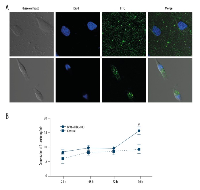
Figure 3.
MV uptake by HBL-100 mammary epithelial cells and consequent change in β-casein secretion. (A) Confocal microscopy images of HBL-100 cells (nuclei stained blue by DAPI) exposed to MVs labeled with rat anti-human CD63 monoclonal antibody as a primary antibody and rabbit anti-rat FITC as a secondary antibody (green fluorescence). Initially, the labeled MVs were observed as green spots around the cells (upper panel), whereas MVs were found inside the HBL-100 cells after 4 hours (lower panel). # P<0.05: MVs+HBL-100 group vs. control group. (B) β-casein concentration in the HBL-100 supernatant after 24, 48, 72, and 96 hours of exposure to umbilical cord blood-derived MVs.

