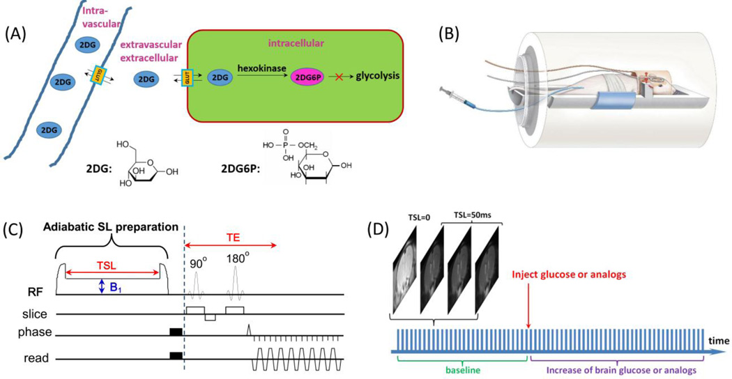
Fig. 1. Experimental setup and protocol for 2DG-CESL study.
(A) A schematic illustration of the transport and metabolism of 2DG in brain. The injected 2DG in the intravascular space is transported by glucose transporters across the blood brain barrier to the extravascular-extracellular space and followed by uptake into the intracellular space where it is phosphorylated into 2DG6P. Both 2DG and 2DG6P have hydroxyl groups which rapidly exchange with water protons and can be detected by the CESL technique. (B) A schematic illustration of our animal-experiment setup shows a combination of volume coil transmit and surface coil receive, and the 2DG is injected through a venous line. (C) The pulse sequence for chemical exchange-sensitive spin-lock (CESL) MRI used in this study contains an adiabatic spin-lock (SL) preparation followed by spin-echo echo planar imaging for imaging acquisition. B1: spin-lock power level, TSL: spin-lock time, TE: echo time. (D) Each glucoCESL MRI experiment was performed by repeated acquisitions of four images (one TSL = 0 ms and three TSL = 50 ms) before and after injection of 2DG or mannitol.
A time series of R1ρ (=1/T1ρ) was generated from data with two TSL values.

