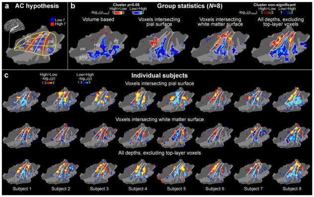
Figure 4.
AC high and low frequency regions identified using high-resolution 7T fMRI sampled across cortical depths. (a) A human AC model hypothesis shown on a flattened patch of left superior temporal cortex (for details, see Fig. 1). (b) Comparison of group analysis results using different analysis approaches. The dotted lines correspond to the main frequency gradients shown in the hypothetical model. An improvement of sensitivity and consistency with the hypothesis is observed when the signals from the pial voxels are excluded. This could reflect improved correspondence of results across the individual subjects. Poorest results are achieved with volume-based analyses (leftmost panel). The main effect of center frequency (100, 240, and 577 Hz vs. 1386, 3330, and 8000 Hz) is shown to help comparisons of frequency-sensitivity regions across subjects. The opaque color scale refers to post-hoc corrected (cluster-based Monte Carlo simulation tests, p<0.05) and the transparent color scale to uncorrected random-effects general linear model (GLM) results. For clarity, the boundaries of the clusters surviving the post-hoc correction have been marked with a white trace. (c) Individual fMRI results on flattened standard-brain AC patches. The upper row shows the data from the voxels intersecting the pial surface, the middle row those intersecting the white matter, and the lowest row shows cortical data with signals from the top layers excluded. Notably, the exclusion of signals from top layers improves the consistency of findings with the hypothesized model in most subjects (e.g., Subj. 8). Another important point to note is the relative weakness of individual-level observations in the white-matter surface: Despite this relative signal weakness, the group level result was statistically stronger than that from the pial surface, in line with our interpretation that the exclusion of pial voxels greatly improves the anatomical consistency of functional results across individual subjects.

