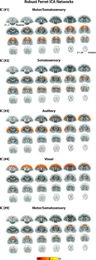
Figure 1.
Ferret gICA networks. IC[#1] Motor/somatosensory network identified from the gICA analysis. Connectivity maps included primary motor cortex (M1 located on the PSG), premotor cortex (PM on ASG), primary somatosensory cortex (S1 on PSG and CNG), higher order somatosensory cortex (tertiary somatosensory area (S3 on SSG), and basal ganglia (caudate nucleus (NC)). Connectivity maps are overlaid onto T2-anatomical images with red-yellow color encoding using a 1.96 < Z-score < 12 threshold (see colorbar); same conventions for ICs #2–4. IC[#2] Somatosensory network identified from the gICA analysis. Connectivity maps included the brain regions primary somatosensory cortex (S1 on PSG and CNG), multisensory cortex (MRSS on medial bank of rostral suprasylvian sulcus), posterior parietal cortex (PPr on SSG) and higher order somatosensory cortex (tertiary somatosensory area (S3 on SSG), and functionally unidentified cortex rostrally adjacent to S1 (on PSG, pro-PSG, and CNG). IC[#3] Auditory network identified from the gICA analysis. Connectivity maps included all primary and higher order auditory cortex fields, and adjacent multisensory and higher order visual cortex fields (summarized, see List 1 for all included regions). IC[#4] Visual network identified from the gICA analysis. Connectivity maps included the brain regions primary visual cortex (area 17), secondary and higher order visual cortex (area 18/19 on LG; area 21 on SSG; ALMS on SSG; ALLS on MEG; suprasylvian cortex (SSy), posterior parietal cortex (PPc on SSG and LG), postsplenial cortex (PSC), functionally undefined cortex (presumed higher order auditory cortex on vPEG) and basal ganglia (caudate nucleus (NC)). IC[#9] Motor/somatosensory network identified from the gICA analysis. Connectivity maps included the brain regions primary motor cortex (M1 on PSG), premotor cortex (PM on ASG), and higher order somatosensory cortex (tertiary somatosensory area (S3 on LG).

