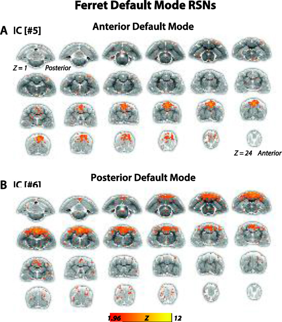
Figure 2.
Anterior and posterior default mode networks of the ferret brain. (A).Putative anterior default mode network (IC[#5]) identified from the gICA analysis. Connectivity maps included the brain regions medial PFC, medial dPFC, and PM. Connectivity maps are overlaid onto T2-anatomical images with red-yellow color encoding using a 1.96 < Z-score < 12 threshold (see colorbar). (B).Putative posterior default mode network (IC[#6]) identified from the gICA analysis. Connectivity maps included the brain regions posterior parietal cortex, posterior cingulate cortex, S3, MRSS and M1.

