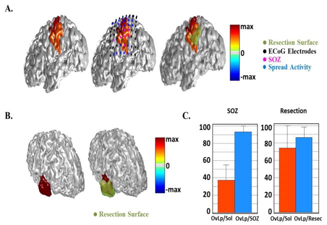
Fig. 11.
Source extent estimation results in all patients. (A) Estimated results by IRES in a parietal epilepsy patient compared with SOZ determined from the intracranial recordings (middle) and surgical resection (right). (B) Estimation results of source extent computed by VIRES in another temporal epilepsy patient compared with surgical resection. (C) Summary of quantitative results of the source extent estimation by calculating the area overlapping of the estimated source with SOZ and resection. The overlap area is normalized by either the solution area or resection/SOZ area.

