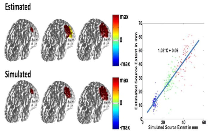
Fig. 4.
Simulation results. In the left panel three different source sizes were simulated with extents of 10 mm, 20 mm and 30 mm (lower row). White Gaussian noise was added and the inverse was solved using the proposed method. The results are shown in the top row. The same procedure was repeated for random locations over the cortex. The extent of the estimated source is compared to that of the simulated source in the right panel. The SNR is 20 dB.

