
FIGURE 5.
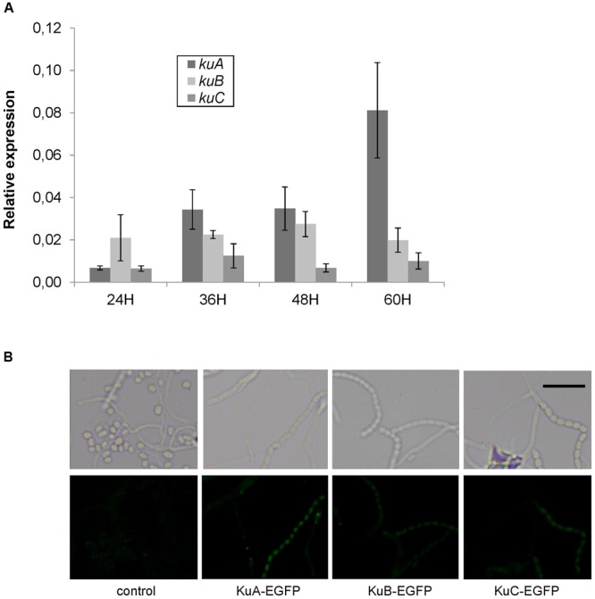
Growth-dependent and cell-type specific expression of NHEJ-like genes. (A) Expression level of S. ambofaciens ATCC23877 ku genes relative to hrdB after 24, 36, 48, and 60 h growth on SFM plates at 30°C. Error bars represent standard error. Expression of kuA after 60 h growth is significantly higher than that at 24 h. Furthermore, kuA expression at 60 h growth is significantly different than that of kuB and kuC (t-test with Bonferroni correction, α = 0.05). (B) Strains expressing EGFP fusion with KuA, KuB, or KuC were inoculated in the acute-angle junction of standard-sized microscope coverslips inserted at 45° in SFM agar, and grown for 48 h before observations by fluorescent microscopy. The control strain does not contain any GFP gene. Representative examples of spores or aerial mycelium are shown as a contrast phase image (upper panels) and as EGFP fluorescent channel (lower panels). The weak fluorescent foci observed mainly in mycelia were readily detected in the control strain and are not likely to correspond to GFP signal. The scale is represented by a 5 μm size bar.