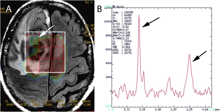
Figure 3.
(a) An axial plane of a fluid-attenuated inversion-recovery sequence is illustrating a high-grade glioma; a selected voxel on the grid MR spectroscopy (MRS) is pointing an area of solid tumour (white arrow). (b) In the MRS spectrum of a tumour region, the major increase of the Cho and LL peaks (black arrows) is evident.

