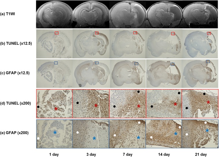
Figure 5.
Colocalization of hollow manganese oxide nanoparticle (HMON)-enhanced MR images and immunohistochemical staining in five pups at each time point: (a) contrast-enhanced T1 weighted images (T1WI) are showing no enhancement at 1 day. HMON enhancement is observed in a part of the injured hemisphere from 3 to 21 days. (b) Corresponding histologic slices with terminal deoxynucleotidyl transferase-mediated dUTP-biotin nick end-labelling (TUNEL) (original magnification, ×12.5) at 1 day are showing a weakly positive staining. TUNEL-positive cells in the injured hemisphere are increasing at 3 days and decreasing gradually by 21 days. (c) Corresponding slices with glial fibrillary acidic protein (GFAP) (×12.5) are showing increased areas of GFAP-positive cells from 1 day to 21 days. (d, e) Magnified slices of TUNEL (d) (red box) (×200) and GFAP (e) (blue box) (×200) are showing negative correlations between the positive areas of TUNEL staining and those of GFAP staining. The areas of TUNEL-positive cells (red asterisks) are negative or weakly positive for GFAP staining (blue asterisks), and the areas of GFAP-positive cells (white dots) are also showing sparse TUNEL-positive cells (black dots).

