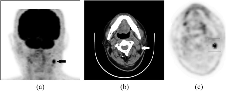
Figure 1.
Fludeoxyglucose (FDG) positron emission tomography (PET)/CT images of a 72-year-old male patient with nasopharyngeal carcinoma who underwent concurrent chemoradiotherapy: focally increased FDG uptake is well visualized in the left neck on the maximum-intensity projection image (a) (black arrow) and the enlarged cervical lymph node (LN) located at the left neck Level II is shown on the transverse CT image (b) (white arrow). Automatic volume of interest (VOI) using an isocontour threshold method was placed over the LN. Segmented VOIs are shown on the transverse PET image (c).

