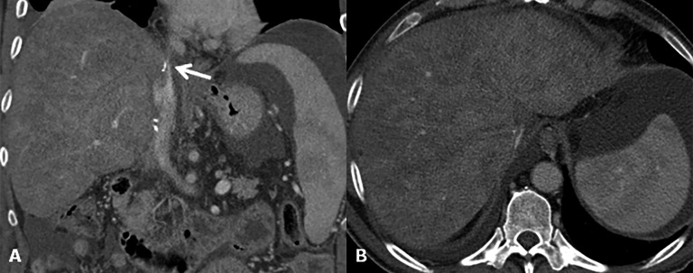
Figure 12.
Outflow obstruction. (a) Coronal CT image after contrast agent injection reveals a supraanastomotic stenosis of the inferior vena cava (arrow). To note on the axial image (b) secondary findings including hepatomegaly, ascites and signs of Budd–Chiari syndrome (liver mosaic pattern perfusion).

