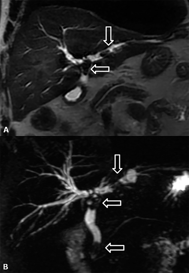
Figure 16.
Lithiasis of the bile ducts. (a, b) Coronal T2 weighted image and maximum intensity projections of three-dimensional thin-slab fast spin-echo T2 weighted image show the dilation of both intra- and extrahepatic biliary tracts with the presence of three stones (arrows) at the level of hepatic bifurcation, in the left hepatic duct and in juxtapapillary site.

