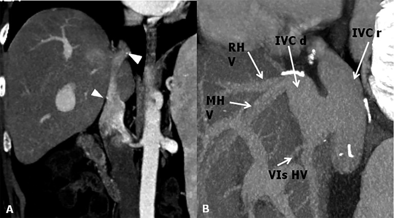
Figure 4.
Inferior vena cava (IVC) anastomoses. (a) CT post-contrast image shows end-to-end anastomosis. To note the surgical clips at the superior and inferior anastomotic sites (arrowheads). (b) Maximum intensity projections sagittal shows a cavoplasty outflow connection, where the graft of the IVC is patched directly onto an incised recipient IVC. IVC d, IVC donor; IVC r, IVC recipient; MHV, mid hepatic vein; RHV, right hepatic vein; VIs HV, hepatic vein for the sixth segment.

