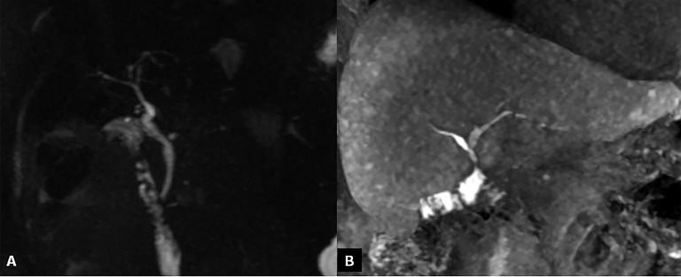
Figure 5.
Bile duct anastomoses. (a) Maximum intensity projections reconstruction of three-dimensional thin-slab fast spin-echo T2 weighted images show a regular hepaticocholedochostomy. (b) Coronal MR contrast-enhanced image obtained after intravenous administration of a hepatobiliary contrast agent (gadoxetic acid); cholangiography shows a normal hepaticojejunostomy.

