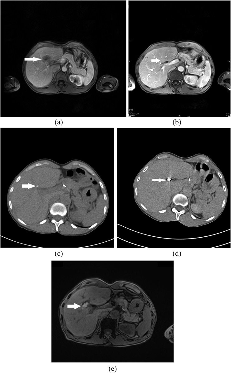
Figure 1.
A 46-year-old female with liver metastases of colorectal cancer (T3N1M1) and morphological features of complete response after treatment with transarterial embolization and microwave ablation (MWA). (a) Pre-treatment contrast-enhanced axial MRI scan shows metastatic liver lesion (arrow) in segment IV. (b) Post-treatment contrast-enhanced axial T1 weighted MR image after three sessions of chemoembolization shows partial response of liver metastases (arrow). (c) CT scan after selective transarterial embolization shows lower degree of Lipiodol retention in metastatic lesion in segment IV (arrow). (d) CT image obtained during MWA shows a microwave antenna positioned in the tumour (arrow). (e) Transverse contrast-enhanced T1 weighted MR image obtained 24 h after MWA demonstrates ablated volume (arrow). Area of necrosis is larger than original lesion.

