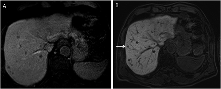
Figure 7.
(a) Pre-contrast T1 MRI fat-suppressed image demonstrating multiple hypodense lesions concerning metastases in a patient with uveal melanoma. (b) Post-contrast T1 MRI hepatobiliary-phase image demonstrating several more lesions in addition to the previously seen lesions. One of the smaller lesions, seen only on the hepatobiliary phase, is indicated by an arrow.

