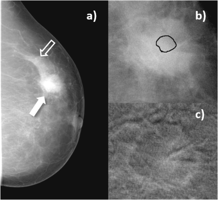
Figure 4.
Craniocaudal images of Patient 4, invasive ductal carcinoma. (a) Mammogram acquired before contrast-enhanced digital mammography procedure. Solid arrow indicates the lesion, open arrow signals a region of normal breast parenchyma; (b) enlarged view of the lesion before contrast medium (CM) injection, with the lesion region of interest drawn by the radiologist; (c) subtracted image of the same region as in (b), 3 min after start of CM injection.

