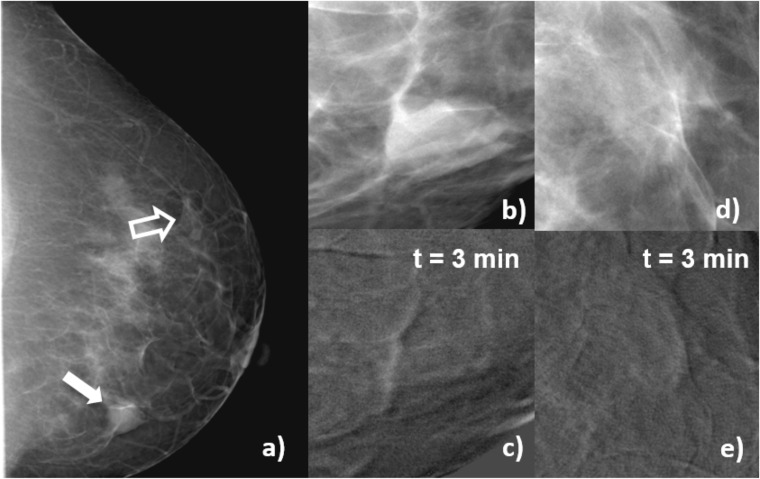
Figure 6.
Craniocaudal images of Patient 3, fibroadenoma. (a) Mammogram acquired before contrast-enhanced digital mammography procedure, the solid arrow indicates the lesion, and open arrow signals a region of normal glandular tissue; (b) enlarged view of a processed (for presentation) image of the lesion before contrast medium (CM) injection; (c) subtracted image of the same region as (b) 3 min after the start of CM injection; (d) enlarged view of a processed (for presentation) image of the glandular tissue before CM injection; (e) subtracted image of the same region as (d) 3 min after the start of CM injection. In the subtracted images, uptake at the lesion region (c) is lower than at the glandular tissue (e), and the contrast is numerically negative. This patient was candidate to a biopsy due to risks factors.

