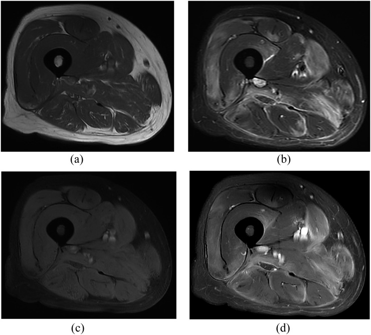
Figure 4.
Corresponding MR images through the mid-thigh level in the same patient as in Figure 3 with polymyositis: (a, b) axial T1 weighted [repetition time (TR)/echo time (TE) = 485/15 ms in (a)] and short tau inversion-recovery [TR/TE = 10,469/65 ms in (b)] MR images are demonstrating multifocal feathery high-signal-intensity infiltration of the entire compartments of the thigh muscles. There is thickening of the subcutaneous fat and skin of posteromedial thigh. (c, d) The axial fat-suppressed, pre-contrast-enhanced and contrast-enhanced T1 weighted images (TR/TE = 632/15 ms) are showing patchy enhancement.

