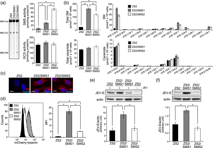
Figure 4. SMS1, but not SMS2, is required for infection and attachment of JEV.
SMS DKO tMEFs stably expressing empty vector (ZS2), mouse SMS1 (ZS2/SMS1), or SMS2 (ZS2/SMS2) were prepared. (a) In vitro SMS and GCS activities were measured in lysate (100 μg protein) of cells using C6-NBD-ceramide as a substrate as described in the Materials and Methods. The values are the mean ± SD (n = 3). *P < 0.005. (b) Measurements of SM and ceramide were performed by LC-MS/MS. The value was mean ± SD (n = 3). *P < 0.005. (c) SM was detected with MBP-lysenin. Nuclei were stained with Hoechst 33342. Scale bars, 10 μm. (d) Membrane SM was detected with mCherry-lysenin and flowcytometer. Mean fluorescence intensity (MFI) was quantified with Kaluza software. (e) To assessed the JEV infection, cells were infected with JEV (MOI = 0.1) for 1 h, washed, and cultured for 48 h. JEV-E and actin were detected by immunoblotting and quantified. (f) For JEV attachment, cells were treated for 15 min with JEV-containing medium derived from Vero cells culture infected with JEV (MOI = 1) for 48 h. Then, JEV-E and actin were detected by western blot analysis and quantified. The presented values are the mean ± SD (n = 3). *P < 0.005. Full-length blots are presented in Supplementary Figure S5.

