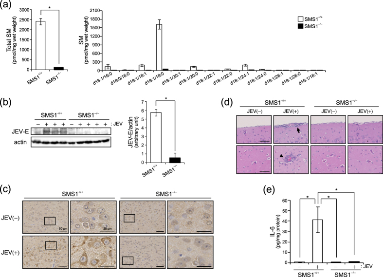
Figure 5. JEV infection of brains was decreased in SMS1 deficient mice.
(a) SM levels were measured by LC-MS/MS in the brains of wild type (SMS1+/+) and SMS1 knockout (SMS1−/−) mice (n = 5). (b–d) SMS+/+ and SMS−/− mice were intraperitoneally injected with JEV (1 × 10⁴ pfu/mouse) (n = 4) and were killed after 13 days. However, one JEV-injected SMS+/+ mouse was dead at 11 day. (b) JEV-E and actin were detected by western blot in brain lysates and quantified. The presented values are the mean ± SD (n = 3 [SMS1+/+] and 4 [SMS1−/−]). *P < 0.005. Full-length blots are presented in Supplementary Figure S6. (c) Immunohistochemistry using anti-JEV-E antibody and BCA staining in brain sections. (d) Haematoxylin and eosin staining of sections of brain. The arrow and arrow head indicate meningitis and leukocyte infiltration, respectively. Scale bars, 50 μm. (e) ELISA of IL-6 concentration in brain lysate. The values are the mean ± SD. *P < 0.005.

