
Figure 7. Fine tuning of Rac1 and RhoA altered cuspal shape through the remolding of cellular geometry in the IDE.
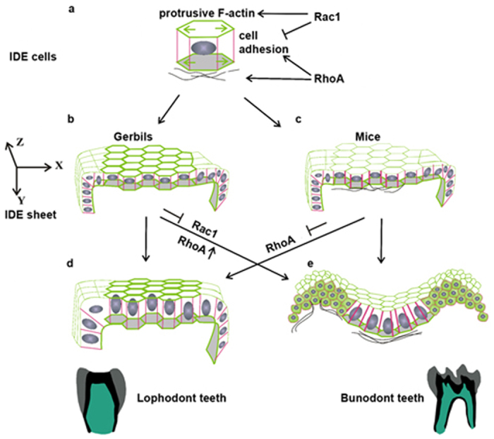
(a) A proposed model showing Rac1 and RhoA control of the cytoskeleton (F-actin), AJs (E-cad), and FN deposition in IDE cells. (b,c) Different distributions of F-actin, E-cad, and FN are present in IDE cells of gerbils and mice. X, Y, Z indicate the axes in three-dimensional space. (d,e) Schematic IDE sheets show that Rac1 and RhoA participate in tissue morphogenesis and cuspal shapes by changing the cell shapes, sizes, and FN assembly. IDE, inner dental epithelium.