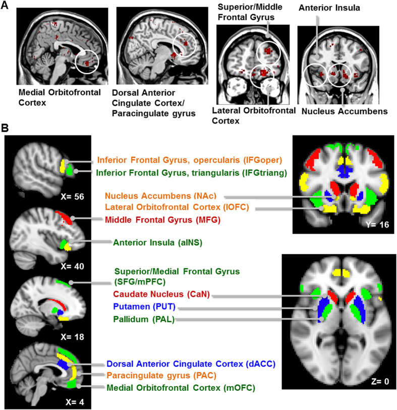
Figure 2. The risk-related network derived from an imaging meta-analysis implemented using Neurosynth (see SI Methods for detailed procedures).
The network composed of anterior insula, nucleus accumbens, the orbitofrontal cortex, the anterior cingulate gyrus, and the lateral and medial prefrontal cortex (Panel A). The brain regions (i.e., the nodes) of the risk-related network in the rsFC connectome analysis (Panel B).

