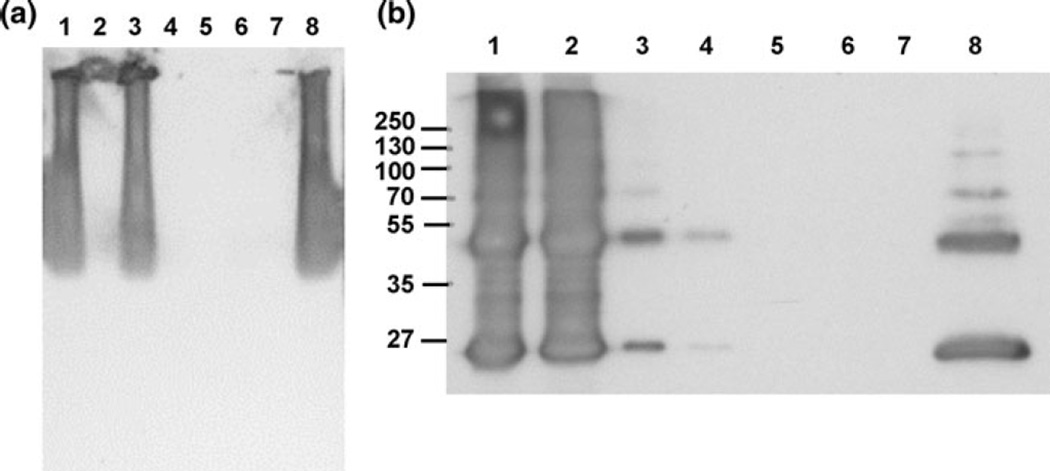
Fig. 11.
a Western blot of the results of the pull-down assay, showing that GXM is washed out in the flow-through and is not retained on the resin. Lane 1–3 flow-through; lane 1 resin control (+GXM), lane 2 bait control (−GXM), lane 3 sample (+GXM), Lane 4–6 eluate; lane 4 resin control, lane 5 bait control, lane 6 sample, lane 7 blank, lane 8 positive control (GXM 1 µg/µl, 15 µg loaded). b Western blot of co-IP showing that App1 elutes in the flow-through (FT) and is not bound to GXM. Lane 1–4 flow-through; lane 1 FT (negative control), lane 2 FT (sample), lane 3 FT after conditioning buffer for negative control, lane 4 FT for sample after conditioning buffer; lane 5 eluate (negative control), lane 6 eluate (sample), lane 7 blank, lane 8 App1 positive control 10 ng

