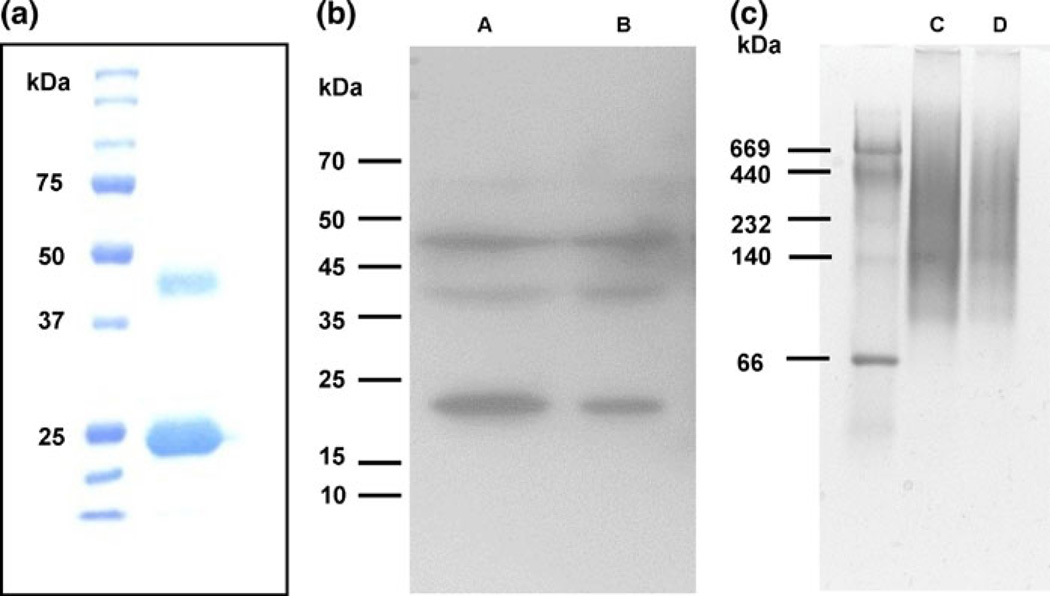
Fig. 2.
a Fifteen percent Coomassie-stained SDS gel of rApp1. b Western blot from 12% SDS–PAGE of total protein extracted from WT, Lane A 20 mg total protein, Lane B 10 mg total protein; 1° antibody: anti-App1 4–6H (1:20), 2° antibody: goat anti-mouse IgG-HRP (1:5,000), 15 min exposure. c Native PAGE (4–20% Tris–HCl gel) of rApp1, Lane C 20 mg, Lane D 10 mg, stained with GelCode Blue

