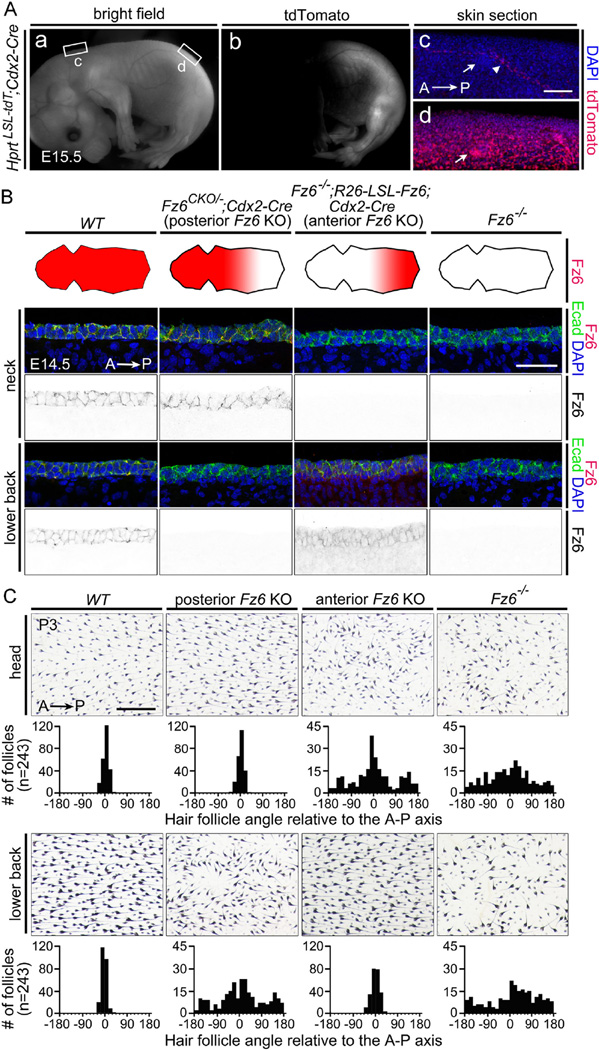
Fig. 2.
Selective deletion of Fz6 in the anterior or posterior of the embryo leads to regional loss of follicle orientation. (A) Cdx2-Cre recombines the Hprt-LSL-tdT reporter throughout the posterior embryo, with the boundary between expressing and non-expressing tissue approximately at the level of the umbilical vessels. Inset rectangles marked (c) and (d) are enlarged to the right. Arrows in panel (c) and (d) point to hair follicles; the arrowhead in (c) points to a blood vessels, the only structures that show Cre-mediated activation of the tdTomato reporter anterior to the mid-abdominal boundary. A, anterior; P, posterior. Scale bar, 100 µm.(B) Elimination of Fz6 in the anterior or posterior halves of the embryo. The four schematics show a flat-mounted dorsal mouse skin with the head to the left. The territory of Fz6 expression is indicated in red. Lower panels, immunostaining for Fz6 and E-cadherin confirms the territorial expression diagrammed above. Scale bar, 50 µm. (C) Follicle orientations in the head (upper panels) and lower back (lower panels) in P3 skin flat mounts from mice with the same four genotypes as shown in panel (B). See Methods section for details on quantification. Scale bar, 0.5 mm.

