Fig. 1.
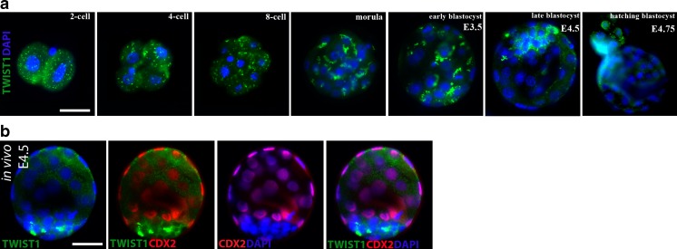
Cellular localization of Twist1 in mouse preimplantation embryos. a Representative images of Twist1 protein during in vitro embryo development (N = 10 embryos per group). Whereas cleavage stage (two- to eight-cell), morula, and early blastocyst stage (E3.5) embryos showed accumulated staining pattern in the cytoplasm, staining was visible in the cytoplasm and perinuclear areas of embryonic cells in ICM layer of late blastocyst at E4.5, and it had been gradually translocated to the nucleus of hatched embryonic cells at E4.75. b Representative images of double-labeled blastocysts obtained in vivo at E4.5 (N = 14 embryos). Twist1 was normally distributed in the cytoplasm and perinuclear areas of embryonic cells in the ICM layer, as shown in the control groups. The green signal indicates positive staining for Twist1, the red signal indicates positive staining for Oct43/4, and the blue signal (DAPI) indicates nuclei of the embryonic cells. Scale bar represents 25 μm

