
Figure 1.
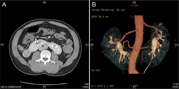
Computed tomography image of a patient with a horseshoe kidney. (A) Axial image demonstrating a tumor involving the right hemi-kidney of the horseshoe kidney (arrow), and a parenchymatous isthmus. (B) 3D reconstruction of the computed tomography image, demonstrating 2 accessory arteries supplying each hemi-kidney. (Color version available online.)