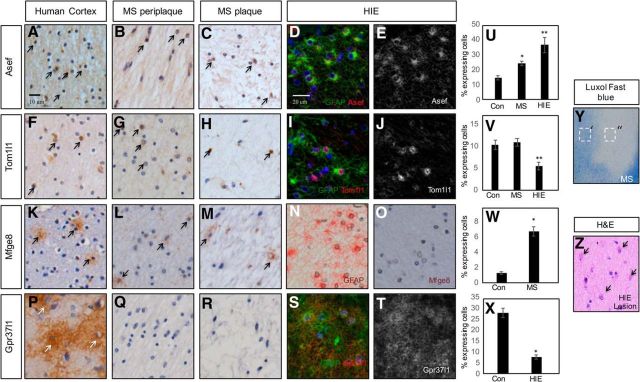
Figure 4.
ASEF, TOM1l1, MFGE8, and GPR37l1 are expressed in human WMI. A–C, IHC demonstrating expression of ASEF in adult human cortex (A) and in human MS plaques (B, C). D, E, Double immunofluorescence in neonatal HIE reveals coexpression of ASEF and GFAP. F–H, IHC demonstrating expression of TOM1l1 in adult human cortex (F) and in human MS plaques (G, H). I, J, Double immunofluorescence in neonatal HIE reveals coexpression of TOM1l1 and GFAP. K–M, IHC demonstrating expression of MFGE8 in adult human cortex (K) and in human MS plaques (L, M). N, O, IHC for GFAP (N) and MFGE8 (O) in neonatal HIE in adjacent section. No expression of Mfge8 is found in HIE with active gliosis. P–R, IHC demonstrating expression of GPR37l1 in adult human cortex (P) and no expression in human MS plaques (Q, R). S, T, Double immunofluorescence in neonatal HIE reveals coexpression of GPR37l1 and GFAP. Arrows denote IHC detection of expression of a given gene. U, Quantification of ASEF expression in MS and HIE; * and **p < 0.0001. V, Quantification of TOM1l1 expression in MS and HIE; * is not significant and **p = 0.005. W, Quantification of MFEG8 expression in MS; *p < 0.0001. X, Quantification of GPR37l1 expression in HIE; * and **p < 0.0001. Y, Luxol fast blue staining of human MS lesion. Dashed boxes denote periplaque (′) or plaque (″) regions. Z, H&E stain on neonatal HIE tissue. Arrows denote reactive gliosis.

