Figure 7.
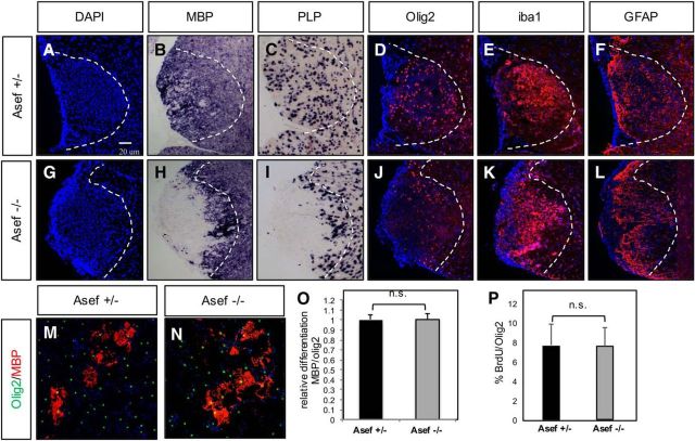
Loss of Asef results in delayed repair after WMI. A–L, Analysis of cellular responses after lysolecithin injury at 10 dpl in the adult spinal cord of Asef+/− (A–F) and Asef−/− (G–L) mice. Asef−/− mice demonstrate reduced expression of mature oligodendrocyte markers MBP and proteolipid protein compared with Asef+/− controls (B, C vs H, I), coupled with a redistribution of Olig2-expressing OPCs within the lesion (D vs J). Expression of reactive astrocyte (F vs L) and microglia (E vs K) markers within the lesion is unaffected. The images are representative of eight mice for each genotype, of which 2–3 sections for each marker were analyzed from each of these mice. M, N, Immunostaining of differentiated OPCs derived from Asef+/− and Asef−/− mice, with markers of mature oligodendrocytes (MBP) and precursors (Olig2). O, Quantification of the number of MBP-expressing cells across three experiments, performed in triplicate, indicates there is no difference in OPC differentiation between these genetic conditions. P, Quantification of the number of Olig2-expressing OPCs labeled with BrdU across three experiments, performed in triplicate, indicates that there is no difference in OPC proliferation between these genetic conditions.

