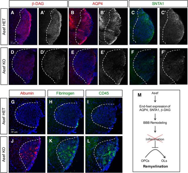
Figure 8.
Impaired reactive astrocyte response in the absence of Asef. A–F, Immunofluorescence demonstrating expression of β-DAG, AQP4, and SNTA1 in Asef+/− (A–C) and Asef−/− (D–F) mice; sections are from adult spinal cord lysolecithin lesions at 10 dpl. Asef−/− mice exhibit decreased expression of β-DAG (A vs D), AQP4 (B vs E), and SNTA1 (C vs F) within lesions. G–L, Immunofluorescence demonstrating expression of albumin, fibrinogen, and CD45 in Asef+/− (G–I) and Asef−/− (J, K) mice at 10 dpl. Asef−/− mice exhibit increased expression of serum/blood proteins albumin and fibrinogen (G, H vs J, K), as well as increased presence of immune cells marked by CD45 (I vs L). The images are representative of four mice for each genotype, of which 2–3 sections for each marker were analyzed from each of these mice. M, Summary of Asef function in reactive astrocytes and BBB remodeling after WMI.

