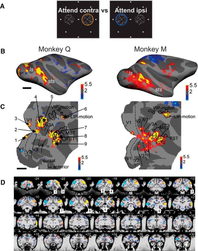
Figure 2.

Spatial attention modulates activity in specific cortical areas of the occipital, temporal, parietal, and frontal lobes. A, Schematic of the two stimulus conditions contrasted in Figure 2: attention to the contralateral RDS (attend contra) versus attention to the ipsilateral RDS (attend ipsi). Attention location is indicated by a colored ring over the cued surface. Significant response enhancements for the attend contralateral versus the attend ipsilateral condition are shown in yellow and red; significant response enhancements to the opposite condition are in blue. B, Statistical parametric maps for the contrast attend contra vs attend ipsi; conventions as in Figure 1D, top. C, Same parametric maps overlaid on flattened posterior hemispheres; conventions as in Figure 1D, bottom; thresholds at p < 0.05, corrected. Left, Numbers point to regions of significant activation shown on coronal slices in D: 1, area V1 lower hemifield; 2, foveal representation; 3, area V1 upper hemifield; 4, area V2; 5, area V3; 6, posterior parietal area LIP; 7, area V4t; 8, area MT; 9, area PITd. D, Parametric maps on coronal slices of high-resolution anatomy, left hemisphere on the right. Cyan and blue indicate higher activity for contrast attend left > attend right; yellow and red indicate higher activity for contrast attend right > attend left.
